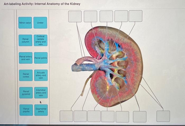
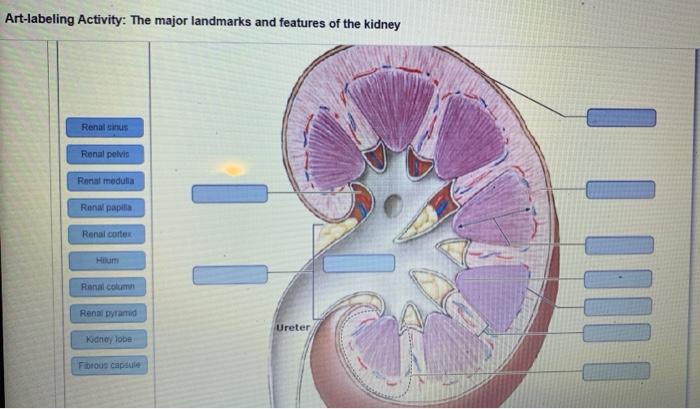
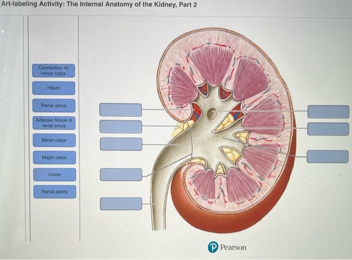
Art-labeling activity: the major landmarks and features of the kidney – Embarking on an art-labeling expedition into the realm of the kidney, this activity unveils the intricate landmarks and features that define this vital organ. By meticulously labeling each anatomical structure, we embark on a journey to unravel the kidney’s complex functions and clinical significance.
Through this immersive exploration, we delve into the kidney’s macroscopic and microscopic architecture, identifying the renal cortex, medulla, and pelvis, as well as the glomerulus, proximal tubule, loop of Henle, and collecting duct. Each structure plays a crucial role in the kidney’s intricate filtration and regulatory processes.
Introduction: Art-labeling Activity: The Major Landmarks And Features Of The Kidney

Art-labeling, also known as anatomical labeling, is a crucial technique in medical education and research. It involves identifying and labeling the major landmarks and features of organs, such as the kidneys, to facilitate a deeper understanding of their anatomy and function.
The kidneys play a vital role in maintaining overall body health by filtering waste products from the blood, regulating fluid and electrolyte balance, and producing hormones. A thorough understanding of the kidney’s anatomy is essential for healthcare professionals to diagnose and treat various kidney diseases.
Major Landmarks of the Kidney
The kidneys are bean-shaped organs located on either side of the spine, just below the rib cage. They are composed of three distinct regions:
- Renal cortex:The outermost layer of the kidney, which contains the glomeruli and proximal tubules.
- Renal medulla:The middle layer of the kidney, which contains the loops of Henle and collecting ducts.
- Renal pelvis:A funnel-shaped structure that collects urine from the collecting ducts and directs it to the ureter.
Microscopic Features of the Kidney
At the microscopic level, the kidney is composed of several specialized structures:
- Glomerulus:A network of tiny blood vessels where blood is filtered to remove waste products.
- Proximal tubule:A coiled tube that reabsorbs essential nutrients and water from the filtrate.
- Loop of Henle:A U-shaped structure that helps maintain the kidney’s concentration gradient.
- Collecting duct:A tube that collects urine from the proximal tubules and loops of Henle.
Clinical Significance of Art-Labeling
Art-labeling plays a critical role in the diagnosis and treatment of kidney diseases. By accurately identifying and labeling the various landmarks and features of the kidney, healthcare professionals can:
- Identify and characterize different types of kidney abnormalities, such as cysts, tumors, and inflammation.
- Assess the severity of kidney damage and monitor disease progression.
- Plan and guide surgical procedures, such as kidney transplants and nephrectomies.
Recent Advancements in Art-Labeling

Advancements in medical imaging technology have significantly improved the accuracy and efficiency of art-labeling. These include:
- High-resolution computed tomography (CT) scans:Provide detailed cross-sectional images of the kidneys, allowing for precise identification of anatomical structures.
- Magnetic resonance imaging (MRI) scans:Offer excellent soft tissue contrast, enabling visualization of the kidneys and their surrounding structures.
- 3D reconstruction techniques:Combine multiple images to create a three-dimensional model of the kidneys, providing a comprehensive view of their anatomy.
Future Directions in Art-Labeling

Future research in art-labeling aims to:
- Develop automated methods for identifying and labeling kidney structures, reducing the need for manual annotation.
- Incorporate artificial intelligence (AI) algorithms to analyze art-labeled images and identify subtle abnormalities that may be missed by the human eye.
- Explore the use of art-labeling in personalized medicine, tailoring treatment plans based on the specific anatomical characteristics of the patient’s kidneys.
Commonly Asked Questions
What is the purpose of art-labeling in kidney anatomy?
Art-labeling allows for precise identification and understanding of the kidney’s various anatomical structures, facilitating a deeper comprehension of their functions and relationships.
How does art-labeling aid in diagnosing kidney diseases?
Art-labeling enables the visualization of abnormal structures or patterns within the kidney, assisting in the identification and characterization of specific kidney diseases.